Ghanaati-Education

"The Role of Jaw Bone Health in Systemic Health"
 

By Prof. Dr. Dr. Dr. Shahram Ghanaati
MD, DDS, PhD

What is Ghanaati-Education?

A short Introduction...

Covered Socket Residuum (CSR) - A Newly Discovered Mechanism of Jaw Bone Healing After Tooth Extraction

For decades, alveolar bone healing after tooth extraction was considered a fully regenerative process. Recent research by Professor Shahram Ghanaati, however, has uncovered a previously unrecognized biological mechanism guiding socket healing.

Using three-dimensional cone-beam computed tomography (CBCT), these radiological studies enabled detailed visualization of post-extraction socket remodeling.

Within six months, significant structural changes were observed: both the buccal and lingual bone plates exhibited inward contraction, resulting in socket volume collapse of up to 55% horizontally and 34% vertically. This collapse represents a natural mechanism by which the body reduces the diameter of the extraction socket from a large “critical-size defect” to a smaller “non–critical-size defect”, enabling spontaneous bone regeneration in the crestal region of the socket.

In the meantime, bone formation predominantly occurs along the peripheral socket walls, while the central region of the socket often remains unmineralized and is radiologically presented as a hypodense area.

As a result, a structure develops that is sealed by a mineralized crestal plate, while its interior remains incompletely ossified—the Covered Socket Residuum (CSR). Radiological analysis demonstrates incomplete internal mineralization. CSR therefore represents a biologically regulated socket-closing process that provides structural stability without achieving complete bone regeneration.

Differentiation from So-Called Cavitations

In the past, similar radiological findings were often interpreted as “cavitations” associated with chronic systemic inflammation. Current evidence, however, clearly differentiates the Covered Socket Residuum (CSR) from these concepts.

Covered Socket Residuum (CSR) is not pathological per se, but represents a physiological, biologically regulated socket-closing mechanism—a programmed adaptation that transforms a post-extraction defect into a stable, partially crestally mineralized structure within the alveolar ridge, detectable by three-dimensional imaging. 


This distinction establishes, for the first time, a scientific separation between disease-related bone alterations and a natural, adaptive healing response within the jawbone.


Health Implications of Hypodense and Hyperdense Jaw Bone Structures Following Tooth Extraction

  • Both hypodense structures—such as the Covered Socket Residuum (CSR)—and hyperdense jaw bone alterations following tooth extraction are a central focus of Professor Ghanaati’s ongoing research, as they may represent metabolically and immunologically active tissue regions.

If such areas persist, they may impair local microcirculation and cellular signaling, potentially sustaining low-grade inflammatory processes that can compromise the long-term success of dental implants. Beyond local effects, chronic micro-inflammatory signaling may also influence systemic immune balance, metabolic regulation, and chronic disease patterns.

Current studies therefore aim to identify the biological and clinical conditions under which hypodense CSR regions either fully mineralize or persist over time, as well as to improve the understanding of the formation, persistence, and clinical relevance of hyperdense jaw bone structures. In parallel, Professor Ghanaati has developed the ARENA-Protocol™, a biologically guided surgical concept designed to prevent CSR formation within the socket and to surgically remove both hypodense and hyperdense, sclerotic jaw bone structures, thereby promoting complete neo-epithelialization of the ridge following augmentation.

The overarching goal is to prevent the development of CSR and, when clinically indicated, to surgically treat hypodense and hyperdense jaw bone structures following tooth extraction, thereby enabling stable bone formation within an inflammation-free microenvironment.


Evidence
  • Radiological Evidence:

    3D imaging shows bone collapse of up to 55% within six months after extraction, with unhealed spaces in the center of the socket (Ghanaati et al., 2025; Bioengineering1, Ghanaati et al., 2025; Bioengineering2)


  • Molecular Evidence:

    Initial data shows that CSR's are "biologically" active, producing inflammatory messengers like RANTES/CCL5, which disturb both local healing and the immune system (Lechner 2013; Ghanaati et al., 2025; Biomedicines)


  • Microbiological Evidence:

    Harmful bacteria (including the "red complex" pathogens linked to gum disease) hide deep in the jawbone and can only be removed by specific surgical techniques like decortication
    (Ongoing clinical studies)


  • Histological  Evidence:

     Microscopic analysis jawbone samples from patients with chronic facial pain revealed areas of ischemic osteonecrosis — dead or poorly perfused bone tissue — often accompanied by chronic inflammation.  These findings confirm that hidden, low-grade jawbone infections can exist even when 2D radiographic images appear normal, contributing to persistent neuralgic pain.  (Bouquot et al., 1992; Oral Surgery)                        


Based on these insights
Ghanaati-Education has a clear
order in its Vision Statement:

Integrating jaw bone health and its systemic relevance into medical and dental education to ensure the highest standards of clinical practice and patient care

Our Mission...

From Research to Clinical Application –

The "ARENA - Protocol™"

At Ghanaati-Education our mission is based on the ARENA-Protocol™:
The ARENA-Protocol™ (Alveolar Ridge Enhancement via Neo-Epithelialization following Augmentation)
, developed by our founder Professor Shahram Ghanaati, represents a biologically guided surgical concept for the management of extraction sites.

Comprehensive pre-operative diagnostics, including cone-beam computed tomography (CBCT) and three-dimensional visualization, are essential to assess tooth-related bone alterations within the socket walls and potential involvement of adjacent jawbone structures.

The protocol begins with atraumatic tooth extraction followed by controlled mucoperiosteal flap elevation, providing direct access to sclerotic socket walls and deeper necrotic or sclerotic jawbone regions. Targeted necrectomy and decortication are then performed using rotary or piezosurgical techniques to expose vital, well-vascularized bone as the foundation for biological regeneration.

Adjuvant decontamination using laser and/or ozone therapy supports the reduction of microbial and inflammatory load at the surgical site. To maintain ridge volume and guide regeneration, a biologized bone substitute enriched with autologous blood concentrates and/or additional biologics—such as hyaluronic acid and its derivatives—is applied within the socket (ridge preservation) and along adjacent ridge segments (ridge augmentation). This approach establishes a biologically active matrix that promotes osteogenesis and angiogenesis enabling predictable regeneration and structural reconstruction of the ridge following tooth extraction.

Systemic patient-related factors, including adequate hydration, a balanced nutrient-dense diet, and sufficient intake of essential micronutrients, further support healing by optimizing cellular metabolism, immune regulation, and collagen matrix formation.

A PTFE (polytetrafluoroethylene) membrane is mandatory to prevent socket collapse and to promote controlled neo-epithelialization of the socket-associated ridge segments.  This biologically guided healing strategy was previously referred to as the Guided Open Wound Healing (GOWH™) concept.

By restoring physiological bone healing conditions, the ARENA-Protocol™ effectively prevents the development of Covered Socket Residuum (CSR). Furthermore, it supports long-term ridge volume stability by enabling controlled neo-epithelialization of the alveolar ridge. This represents a fundamentally different biological healing mechanism compared with conventional primary, secondary, or tertiary wound healing concepts.

Preventing CSR may also reduce local and potentially systemic inflammatory burden; however, further controlled clinical and translational studies are required to evaluate long-term outcomes and systemic effects.

Clinical Implementation – Translating the CSR Mechanism into Practice

The discovery of the CSR mechanism and the development of the ARENA-Protocol™ has defined a set of clinical parameters that embody the translational philosophy of Professor Ghanaati and his educational approach.

These central parameters form the foundation of biologically guided treatment:

1. Radiological Detection – 3D visualization and assessment of affected areas within the jaw to identify non-mineralized or metabolically active regions.

2. Nutritional and Vitamin Balance – Optimization of nutrient and vitamin intake pre- and postoperatively to ensure biological readiness for healing.

3. Hydration – Maintaining sufficient patient hydration before and after treatment to support microcirculation and cell metabolism.
 
4. Decortication – Comprehensive cleaning and biological activation of the extraction socket to remove the infected tooth associated infected/necrotic socket bone, in order to enhance vascular and osteogenic responses. 

5. Use of Bone Substitute Materials and Blood Concentrates – Careful selection of biomaterials combined with autologous blood concentrates (PRF) to stimulate angiogenesis, osteogenesis, and neo-epithelialization.

6. Systemic Condition of the Patient – Detailed evaluation of overall health, including pre-existing diseases and metabolic or inflammatory conditions, followed by individualized treatment adaptation.

Together, these parameters translate biological insight into evidence-based regenerative strategies that respect both, local bone biology and systemic health.

Ghanaati-Education –
Bridging Research, Practice, and Teaching

Ghanaati Education translates these biological and clinical insights into structured learning and training modules.

Using an innovative three-dimensional visualization approach based on radiological diagnostics (CBCT and CT), hypodense structures—including the Covered Socket Residuum (CSR)—as well as hyperdense jaw bone alterations following tooth extraction can be reliably detected. The application of ARENA-Protocol™ as an innovative regenerative and reconstructive surgical concept enables jawbone areas to be revitalized.
To generate robust scientific data, each patient’s medical condition is systematically assessed using a comprehensive medical questionnaire developed by Professor Ghanaati. This evaluation begins prior to surgery and continues for up to one year postoperatively. The resulting data provide valuable insights into the systemic medical effects of ARENA-Protocol™–related procedures, enabling continuous refinement of the approach and ongoing improvement of patient outcomes.

Dentists and Oral Surgeons gain a deep understanding of CSR and ARENA-Protocol™ – from identifying biological healing patterns to implementing them clinically.
This integrative approach unites scientific evidence, surgical precision, and systemic health awareness, forming the foundation of modern biologically guided regenerative dentistry and surgery.

To date, more than 100 dental professionals worldwide have received structured education in the ARENA-Protocol® through Professor Ghanaati’s Maxi Residency Program at Tufts University School of Dental Medicine, in collaboration with the Academy of Medical and Oral Regeneration.
In addition, selected dental centers have received certification as ARENA-Protocol® Centers after fulfilling defined educational, clinical, and quality-related criteria, thereby committing to standardized implementation and the highest standards of patient care.

A New Era of Health Focused Guided Healing 

Together, the recognition of the Covered Socket Residuum (CSR) and the application of the ARENA-Protocol™ represent a paradigm shift in how jaw bone healing within the tooth socket is understood, taught, and clinically practiced.

They redefine the post-extraction process not as a binary state of “healed” or “unhealed,” but as a dynamic and biologically variable adaptation.

By integrating research, clinical application, and education, this concept fosters a future in which jaw bone healing is not merely observed, but consciously guided—enabling predictable regeneration, sustainable health, and a biologically intelligent approach to oral surgery.

The Founder

Professor Shahram Ghanaati
MD, DDS, PhD


Professor Shahram Ghanaati is a board-certified oral and maxillofacial surgeon with additional specialization in plastic surgery. He serves as Deputy Director and Senior Chief Consultant of the Department of Oral, Cranio-Maxillofacial and Plastic Facial Surgery at Frankfurt University Medical Center, where he also directs the Head and Neck Cancer Center and the Wound Center. He is internationally recognized for his pioneering research in biomaterials, tissue engineering, and biologics, and for translating these innovations into clinical practice worldwide.

From the beginning of his career, Professor Ghanaati has combined excellence in clinical practice, research, and teaching. Since joining Goethe University Frankfurt in 2007, he has founded the FORM-Lab for biomaterial research and tissue engineering, mentored countless researchers, and established a study unit dedicated to clinical trials in regenerative therapies. Through this work, he has built one of the world’s foremost centers for clinically oriented biomaterial research, translating basic science into everyday clinical practice.
In both his clinical practice and research, Professor Ghanaati focuses on two key areas: regeneration with reconstruction by means of biomaterials and autologous blood concentrates for tissue repair throughout the body. As a leading oncological and reconstructive surgeon, he develops innovative approaches for treating large bone and soft tissue defects following cancer surgery. His work has introduced novel surgical methods, including the ARENA-Protocol for biomaterial-based tissue regeneration and reconstruction of the jaw bone after tooth extraction. 
Since 2010, in collaboration with Dr. Joseph Choukroun, he has advanced the field of autologous blood concentrates, co-developing the Low-Speed Centrifugation Concept (LSCC), which laid the foundation for the AWMF S3-guideline on the clinical use of platelet-rich fibrin in dentistry.

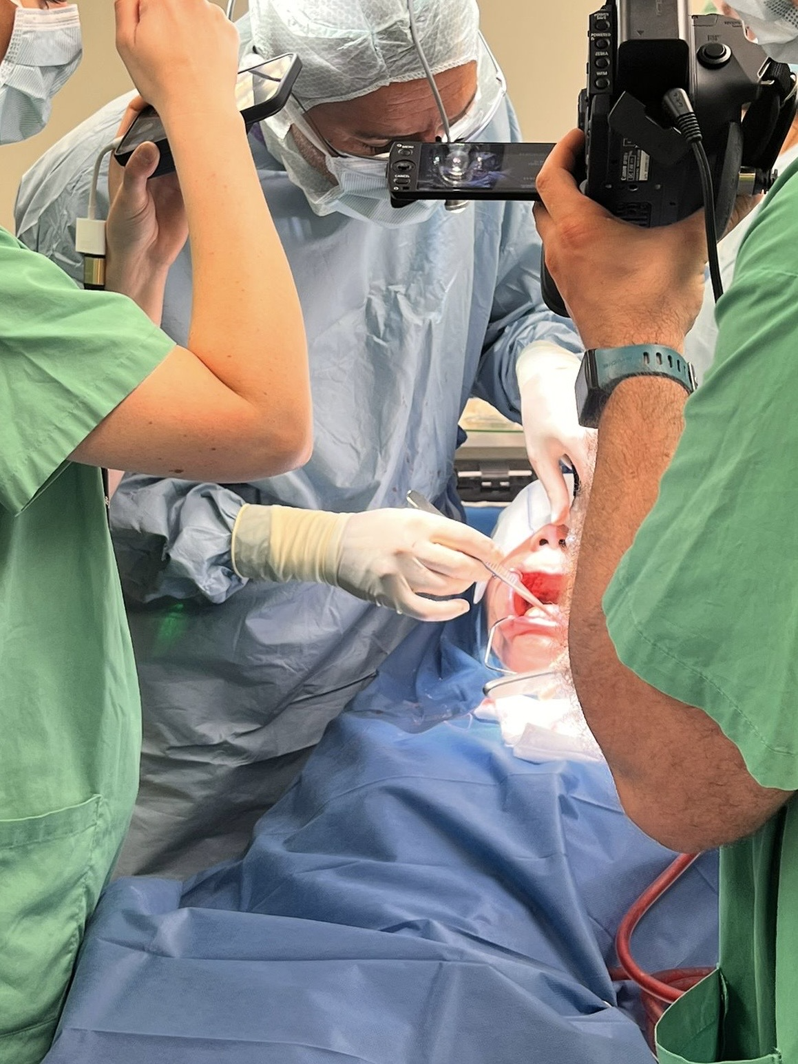
In 2020, Professor Ghanaati founded the Academy for Medical and Oral Regeneration (AMOR), an international platform dedicated to advancing biologics and biomaterials in medicine. AMOR supports basic and clinical research, conducts clinical studies to explore the link between oral health and systemic health, and provides national and international training in GOWH. Building on his extensive experience, he has also pioneered new educational formats for physicians and dentists, integrating cutting-edge science with practical training. To enhance knowledge transfer, he introduced 3D visualization into surgical education and, in 2024, founded Visiogenics and its online platform, ghanaati-education.com, to expand global access to biologic surgical education.
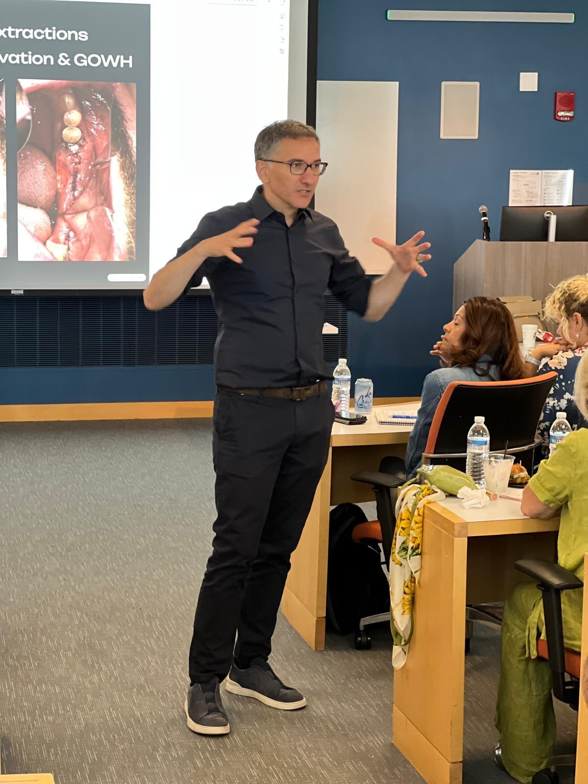
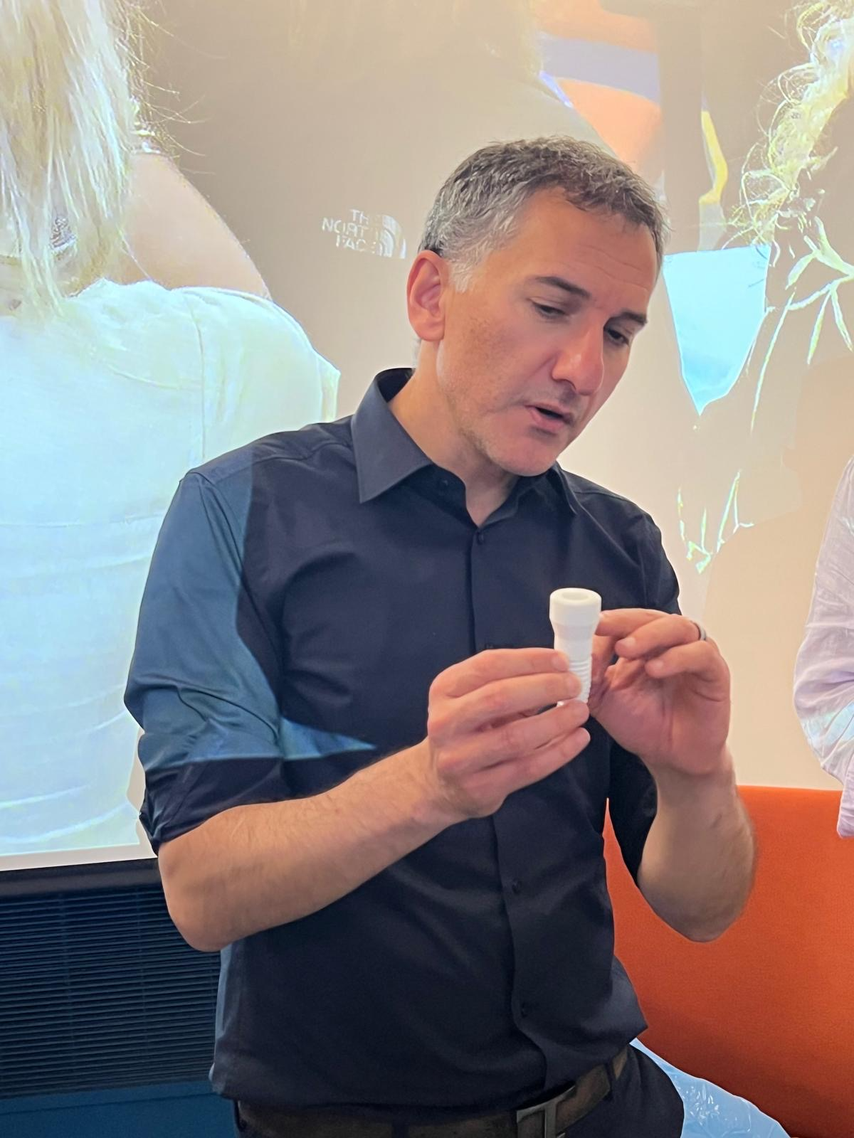
Professor Ghanaati is a highly sought-after speaker, having delivered more than 180 lectures at international congresses and over 250 hands-on courses and workshops worldwide. He has published more than 200 peer-reviewed papers, with an H-Index of 41 and a cumulative impact factor exceeding 640. In addition to serving as a reviewer for numerous journals in regenerative medicine, oral and maxillofacial surgery, and head and neck oncology, he is Senior Associate Editor of the Journal of Oral Implantology (JOI).
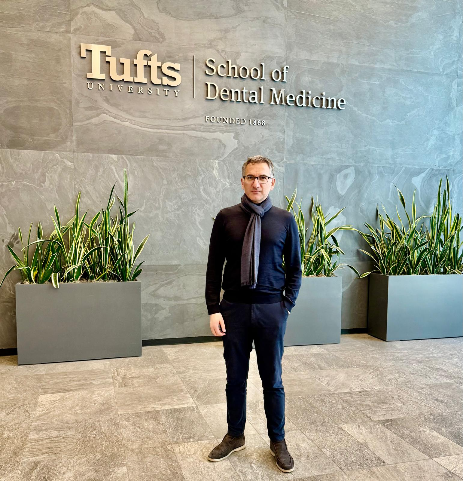
He has received numerous national and international awards for his clinical and scientific contributions and has served as a guest lecturer and adjunct faculty at universities worldwide. Since 2024, he has been Adjunct Instructor at Brooks Rehabilitation College of Healthcare Sciences, Jacksonville University, where he teaches biologic concepts for facial treatment and aesthetics. At Tufts University School of Dental Medicine in Boston, he created the innovative Maxi Residency in Biological Dentistry, Biological Surgery and Implantology based on Guided Open Wound Healing, which combines theory, hands-on training, case presentations, and online modules.